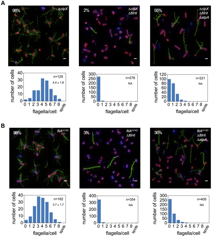
Figure 4. Frequency of flagellar filament formation of a fliHI mutant strain is increased in clpX null and fliAH14D backgrounds.
A deletion in clpX (A) and the more stable fliAH14D variant (B) increase the frequency of flagellar filament formation in a fliHI mutant strain. Flagellar formation is further enhanced by combination with an atpA mutation. Top: A montage of representative fluorescent microscopy images is shown. Flagellar filaments were stained using anti-FliC immunostaining and detected by FITC-coupled secondary antibodies (green), DNA was stained using Hoechst (blue) and cell membranes using FM-64 (red). Scale bar 2 µm. The percentage of cells with at least one filament is presented in the upper left corner. Bottom: Histogram of counted flagellar filaments per cell body. Number of counted cells and average number of filaments per cell +/− standard deviation based on Gaussian non-linear regression analysis is given in the upper right hand corner.

