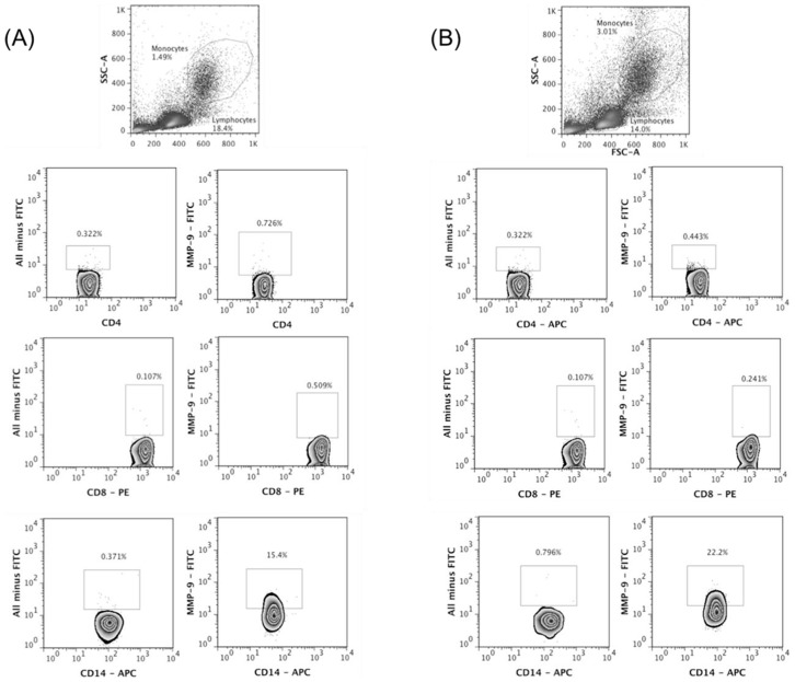
Figure 3. Monocytes are important source of MMP-9 in CL.
PBMC were obtained from healthy subjects (HS) and CL patients and intracellular labeling to MMP-9 was performed ex-vivo. The frequency of CD4+, CD8+ and CD14+ cells positive to MMP-9 was determined by flow cytometry. The gate strategy was done using all minus one staining. A, representative plots from HS (n = 5). B, representative plots from CL patients (n = 5).

