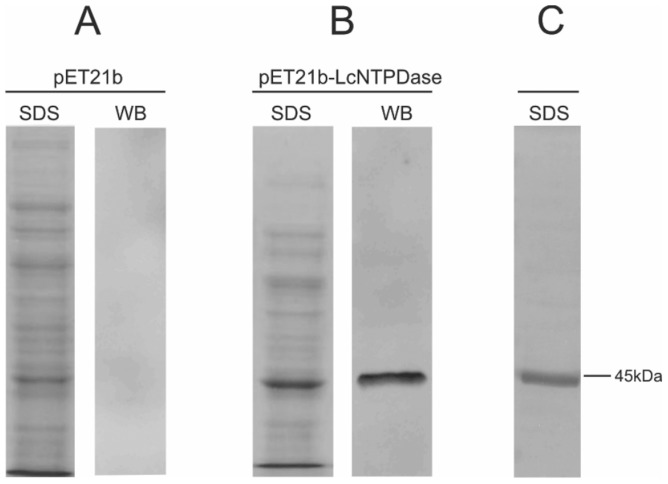
Figure 3. Analyses of expression and purification of rLicNTPDase-2.
(A) Protein extract from E. coli carrying the empty vector pET21b. (B) Protein extract from E. coli carrying the vector pET21b plus rLicNTPDase-2. (C) Purified rLicNTPDase-2 (3 µg) stained by the silver method. SDS lanes indicate samples analyzed after SDS (10%)-PAGE stained with Coomassie blue. WB lanes indicate the same SDS-PAGE samples analyzed by Western blot using anti-His produced in rabbit as primary antibody (1∶4000) and anti-rabbit-IgG conjugated with FITC as secondary antibody (1∶6000). The nitrocellulose membrane was analyzed using an FLA 5100 (Fujifilm) instrument at 475 nm, with a blue filter.

