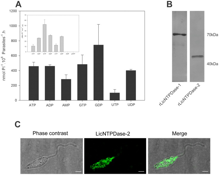
Figure 6. L. infantum chagasi promastigote ecto-nucleotidase activity and surface ENTPDase localization using anti-rLicNTPDase-2.
(A) The promastigote ecto-nucleotidase activity assays were performed in live total promastigotes from log phase growth using different substrates and Mg2+ as cofactor. The activities of recombinant enzyme (rLicNTPDase-2) are shown in the inset box. The SDs represent those of the average of three independent experiments performed in triplicate. The free phosphate released was measured using the malachite green method. (B) Western blotting using anti-rLicNTPDase-2 recognizes both recombinant isoforms rLicNTPDase-1 and rLicNTPDase-2. Purified rLicNTPDase-1 and rLicNTPDase-2 were run in 10% SDS-PAGE and blotted onto a nitrocellulose membrane. The membrane was incubated with purified rabbit polyclonal antibodies to anti-rLicNTPDase-2 (1∶100) as the primary antibody and with anti-rabbit IgG conjugated with FITC (1∶10,000) as the secondary antibody. (C) Distribution of ENTPDases at the surface of L. infantum chagasi promastigotes. Non-permeabilized cells fixed with paraformaldehyde were incubated with anti- rLicNTPDase-2 (1∶50) as primary antibody and with Alexa 488-conjugated goat anti-rabbit IgG (1∶400) as secondary antibody. The glass slides were mounted with Prolong Gold Antifade Reagent (Molecular Probes) and examined by confocal microscopy (Leica, SP5). Bar scale = 2 µm.

