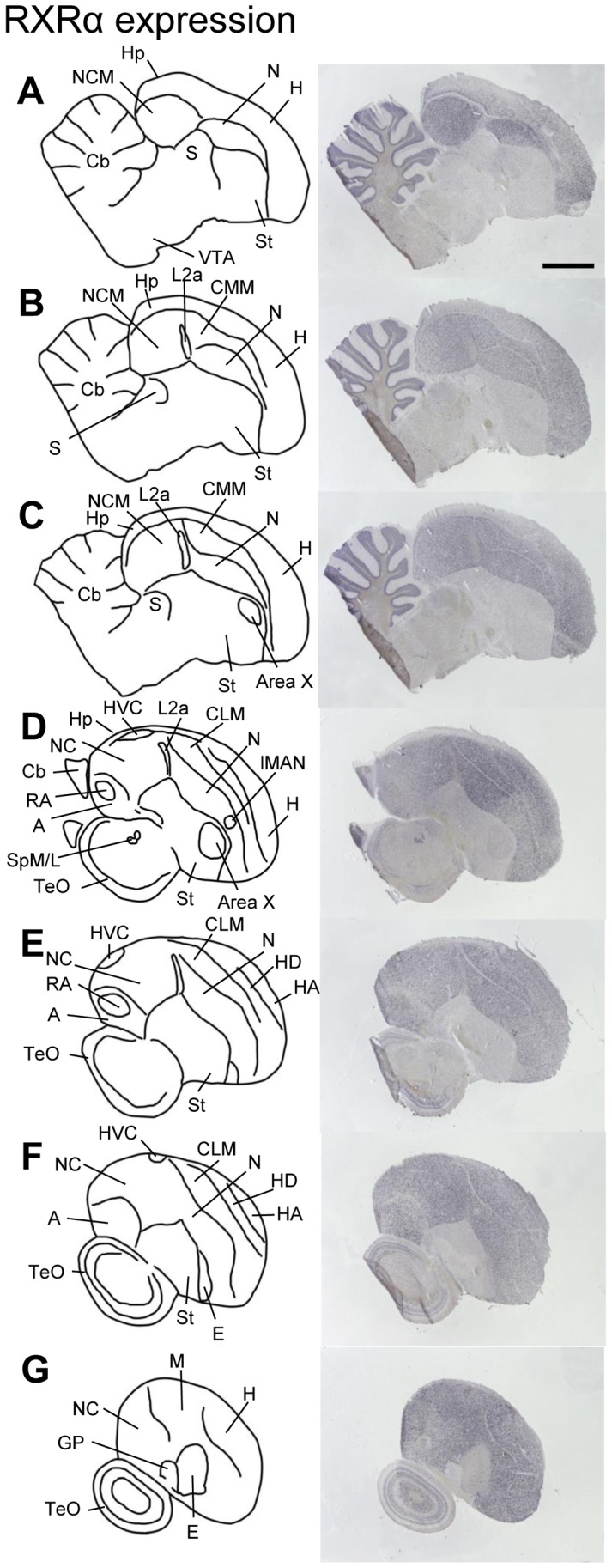
Figure 3. RXRα expression in adult male zebra finch brain.

Drawings on the left depict brain areas and nuclei of serial parasagittal sections. The corresponding sections on the right were processed for in situ hybridization (ISH) for RXRα. For all images, anterior is to the right and dorsal is up; medial to lateral levels are represented from top to bottom. Scale bar = 2 mm (all panels). For abbreviations, see table 1.
