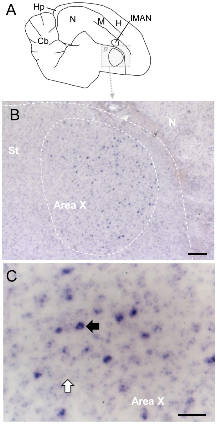
Figure 6. RXRγ expression in Area X of adult male zebra finch.

A: Drawing of a parasagittal section of adult brain, indicating detail area shown in B; anterior is to the right, dorsal is up. For abbreviations see table 1. B: Detail view of Area X and surrounding area in section processed for RXRγ ISH showing sparse labeled cells in Area X. D: High-magnification view of Area X; black and white arrows depict labeled and unlabeled cells, respectively. Scale bars: 2mm in B, 200µm in C, 50µm in D.
