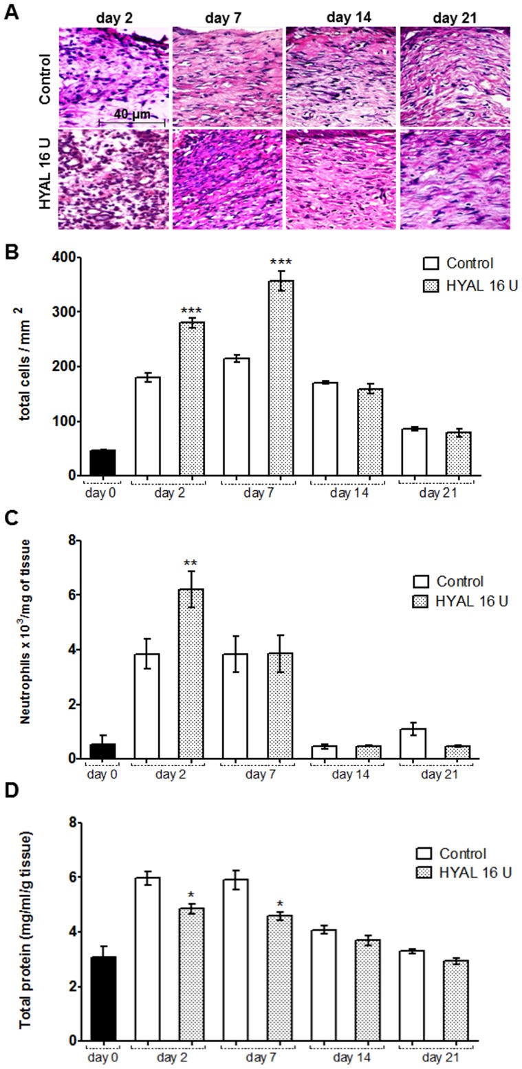
Figure 2. HYAL affects cellular recruitment and edema formation.
Animals were topically treated either with vehicle (control group) or HYAL 16 U daily. Paraffin-wound sections were stained with HE to evaluate the inflammatory infiltrate response by image analysis. (A) The sections were photographed at 400x. The ImageJ software was used to count the inflammatory cells in wound tissue specimens at day 2, 7, 14 and 21 post wounding in at least ten random optic fields per group. (B) Histogram of a quantitative analysis of inflammatory infiltrate counted. (C) Tissue neutrophil accumulation determined by MPO levels in wound biopsies. (D) The total protein content was measured according to Coomassie assay. Values represent mean ± SEM (n = 8 wounds/group), *P<0.05, **P<0.01, ***P<0.001 compared to control group by one-way-ANOVA.

