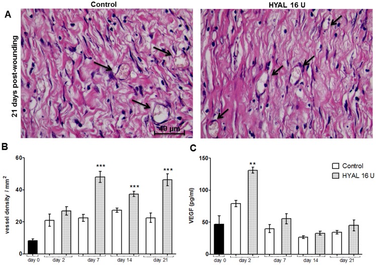
Figure 5. Neovascularization induced by HYAL.
Animals were topically treated either with vehicle (control group) or HYAL 16 U daily. Paraffin-wound sections were stained with HE to evaluate the angiogenic response by image analysis. The sections were photographed at 400x. The ImageJ software was used to count the blood vessels in wound tissue specimens at day 2, 7, 14 and 21 post wounding in at least ten random optic fields per group. (A) Representative photomicrograph of blood vessels (black arrow) in wound tissue specimens at day 21 post-wounding. (B) Histogram of a quantitative analysis of vascular density counted. (C) Vascular endothelial growth factor (VEGF) expression measured in the supernatant of wound tissue homogenate by ELISA. Data represent means ± SEM (n = 8 wounds/group), **P<0.01, ***P<0.001 compared to control group by one-way-ANOVA.

