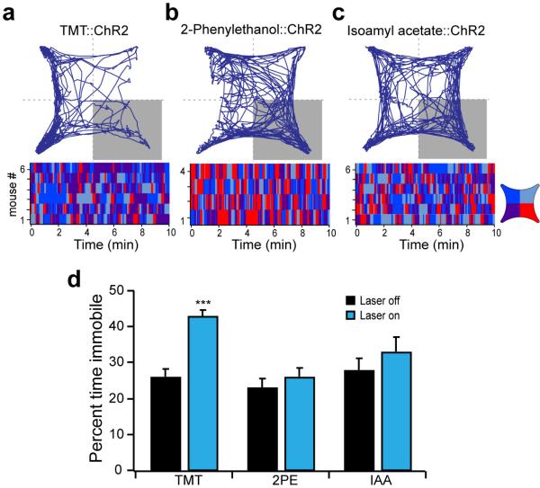
Extended Data Figure 8. Locomotor activity of mice during activation of odor responsive neurons within cortical amygdala.
Mice with odor-driven channelrhodopsin expression were tested in the open field assay where they received pulsed photoactivation upon entrance into the lower right quadrant. a-c, The trajectory graphs (top) show the position of representative animals with ChR2-eYFP in neurons activated by TMT (a), 2-phenylethanol (b) or isoamyl acetate (c). The raster plots (bottom) show quadrant occupancy over time. d, The percent time immobile in the absence and presence of photoactivation. Immobility is defined as velocity less than 1 cm/sec for at least 1 second. a-c, TMT (n=6), 2-phenylethanol (n=4) and isoamyl acetate (n=6); ***P < 0.001 paired t-test comparing with and without laser; error bars show SEM.

