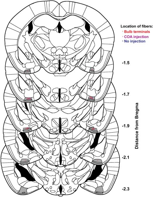
Extended Data Figure 2. Location of optical fibers implanted in cortical amygdala for photoactivation of halorhodopsin.
Schematics show coronal sections throughout most of the region containing cortical amygdala. The posterolateral cortical amygdala is highlighted in gray and the location of bilaterally implanted fibers is indicated.

