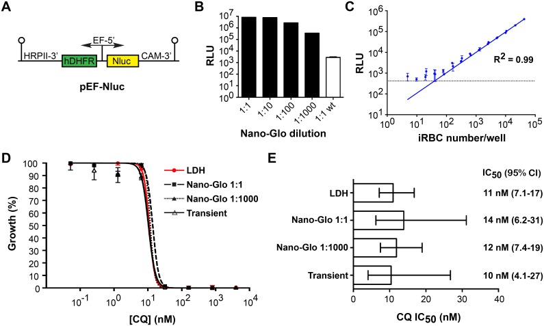
Figure 2. Plasmodium falciparum stably expressing Nluc.
(A) The Nluc gene was cloned in the pEF vector for stable expression in P. falciparum. (B) Aliquots (100 µL) of cultures at 1% hematocrit and 5% parasitemia corresponding to 500,000 P. falciparum infected RBCs transfected with pEF-Nluc were mixed with 1 volume of Nano-Glo Luciferase Assay Reagent and reporter activity measured (1∶1). Nano-Glo Luciferase Assay Reagent was further diluted in 10-fold increments in Luciferase Cell Culture Lysis Reagent and used to determine reporter activity of the same culture. As a negative control, wild type parasites (wt) were mixed 1∶1 with Nano-Glo Luciferase Assay Reagent. (C) Parasites stably transfected with pEF-Nluc were diluted in 2-fold increments in RPMI + RBC maintaining 0.5% hematocrit. For each sample, 10 µL of the culture dilutions were mixed with 40 µL of Nano-Glo diluted 1∶400 in water and measured in the luminometer for 2 s with the gain adjusted 10% below saturation for the brightest sample. The solid line represents the mean RLU after linear regression. The dashed line represents the background +3 standard deviations. (D) Nluc expressing parasites were cultured in varying concentrations of chloroquine (CQ) and their growth determined by the LDH standard method or by measuring reporter activity using Nano-Glo Luciferase Assay Reagent at its standard dilution (1∶1) or diluted 1∶1000 as described in (C). Similarly, wild type parasites were transiently transfected with pPfNluc and their growth determined using Nano-Glo Luciferase Assay Reagent (Transient). IC50 was calculated by non-linear regression and represents the mean of 3 experiments. (E) The IC50s determined in D were plotted with 95% confidence intervals (CI).

