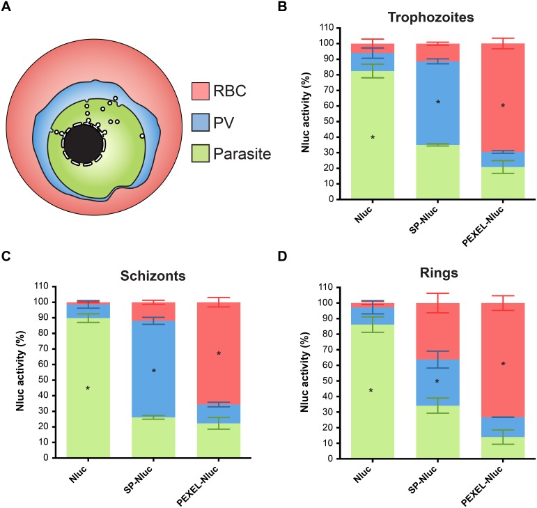
Figure 4. Quantification of Nluc in cellular compartments of infected RBCs.
(A) Schematic of the iRBC’s compartments where RBC represents the exported fraction that is released after Equinatoxin treatment. The PV compartment was then released following treatment with 0.01% saponin and finally, the Parasite fraction was lysed by Nano-Glo Luciferase Assay Reagent. (B) Trophozoite stage parasites transfected with either the original Nluc, secreted SP-Nluc or the exported PEXEL-Nluc fusions were fractionated as shown in (A) and luciferase activity measured. Nluc activities as a percentage of the total for each parasite line are shown and represent the mean of 3 experiments +/−SEM. Similar to (B), (C) Schizonts and (D) Ring stage parasites were also fractionated. Statistical significance (* p<0.05) was determined by 2 way ANOVA test comparing the percentage of reporter activity of each sub-cellular fraction among the 3 cell lines.

