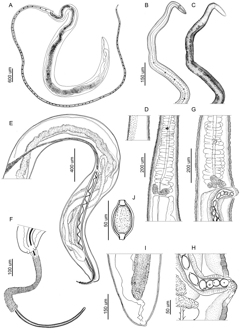
Figure 1. Drawings of Trichuris bainae n. sp.
(A) Complete female specimen. (B) Esophagus, muscular and stichosome portions. (C) Esophagus, muscular and stichosome portions, with bacillary band and cuticular inflations view. (D) Male, esophagus-intestine junction and proximal portion of testis, with bacillary band view. (E) Male, posterior end, spiny spicular sheath, spicule and proximal and distal cloacal tube, lateral view. (F) Male, detail of the posterior extremity, lateral view. (G) Female, esophagus-intestine junction and vulva, lateral view. (H) Female, detail of vulva, lateral view. (I) Female, posterior end, lateral view. (J) Egg.

