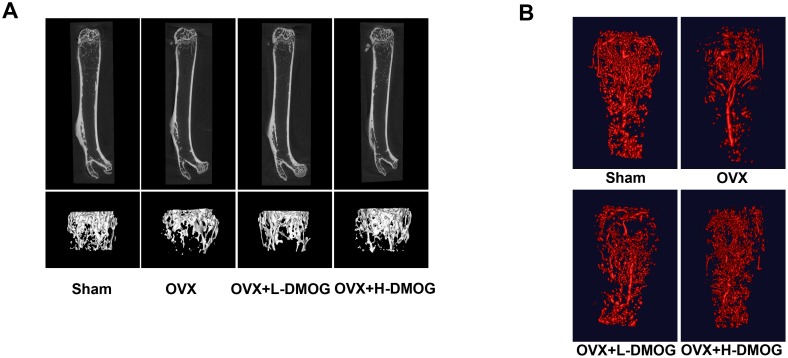
Figure 3. BMD and bone microarchitecture of the trabecular bone in the distal femur.
(A) DMOG abrogated the decrease in BMD and the deterioration in bone microarchitecture induced by OVX, as measured by micro-CT. Morphological analysis of the vasculature within the distal femur from Microfil-perfused mice. (B) OVX decreased the vessel volume at the distal metaphysic of the femur, while DMOG treatment partly restored vessel volumes.

