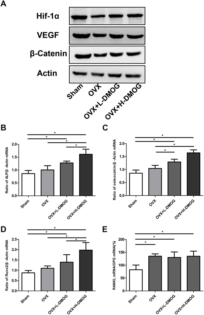
Figure 5. HIF-1α, VEGF, and β-catenin expression in bone samples detected by western blot.
(A) HIF-1α, VEGF, and β-catenin expression was lower in OVX mice than in sham mice. However, DMOG treatment increased HIF-1α, VEGF, and β-catenin expression relative to expression in the OVX group. Effects of DMOG administration on tibial ALP, osteocalcin, RUNX-2, and RANKL/OPG mRNA expression in OVX mice assessed by real-time PCR. (B) ALP/β-actin ratio. (C) Osteocalcin/β-actin ratio. (D) RUNX-2/β-actin ratio. (E) RANKL/OPG/β-actin ratio. P<0.05 for comparisons among the groups designated with an asterisk.

