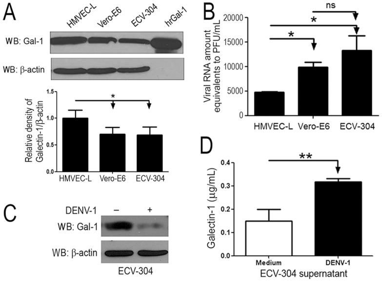
Figure 1. Lower expression of Gal-1 is correlated with higher viral loads produced by DENV-1-infected cells.
(A) Gal-1 expression on HMVEC-L, Vero-E6 and ECV-304 cells was assessed by western blot method and normalized by β-actin endogenous control. The relative density of Gal-1 was determined by ImageJ software. (B) HMVEC-L, Vero-E6 and ECV-304 cells (2.5x10⁴) were incubated with DENV-1 (MOI 0.5) for 72 hours at 37°C. At the end of incubation period, the total amounts of viral RNA in the cell-free supernatants were determined by Real-Time PCR, using a standard curve constructed from DENV-1 RNA purified from 1x10⁷ PFU (PFU: plate formed units). Results are shown as Viral RNA amount equivalents to PFU/ml±SD from 3 independent assays performed in triplicates. (C) ECV-304 cells were inoculated with DENV-1 (MOI 0.5) or only with medium and cultivated for 72 hours at 37°C. Cells were analyzed for Gal-1 expression by western blot assay. (D) Soluble Gal-1 was detected in the supernatants from cell cultures using ELISA method (N = 3). *p<0.01; **p<0.001.

