
Figure 3. Effect of single dose cranial irradiation on circulating endothelial progenitor cell number.
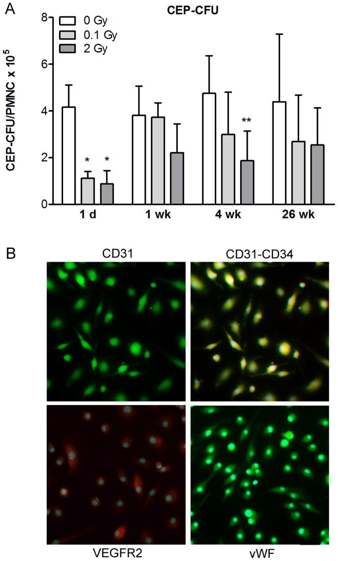
Single head irradiation with 0.1, 2 and 10 Gy doses was performed on 10-week-old adult mice. Circulating endothelial progenitor cell were isolated and cultured from peripheral blood mononuclear cells 1 day, 1, 4 and 26 weeks postirradiation (A). The number of endothelial progenitor cells is presented as colony forming unit/peripheral mononuclear cell ×105/ml blood. Values presented are means ± SD, n = 3–10 from 2–3 separate experiments. Statistical analysis: two-way ANOVA followed by Bonferroni post-test. Statistically significant differences p<0.05 (*) and p<0.01 (**) are indicated compared to sham treated animals examined at the same time point. Representative images of endothelial progenitor cells immunostained for markers are presented in the panels (B): single staining for CD31; double labeling for CD31 and CD34; staining for VEGFR and vWF. CEP-CFU: circulating endothelial progenitor cell-colony forming unit; PMNC: peripheral blood mononuclear cell; CD31; cluster of differentiation 31, CD34; cluster of differentiation 34, VEGFR: vascular endothelial growth factor receptor, vWF: von Willebrand factor. Scale bar: 50 µm.