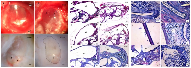
Figure 2. Otitis media is observed in Spag6-deficient mice.
(A–B) Effusion and two bubbles (arrows) could be seen in the mutant mice. (C–D) Some crystals (arrows) were found in a mutant mouse aged 6 months. Fibrous tissue fills the whole middle ear cavity (dotted box). (E–F) At P12 (n = 4, each genotype), the mutant mice did not show otitis media with effusion (OME), some mesenchymal cells (arrows in F) could be observed in the middle ear cavity (ME). (G–H) At P18 (n = 4, each genotype), inflammatory cells began to show in the middle ears of the mutant mice (arrows in H). (I–P) At P180 (n = 1, each genotype), inflammatory cells and effusion were full of the ME(J) and Eustachian tube (ET) (L), Eustachian tube opening (ETO) was blocked by the inflammatory effusions (P). The middle ear epithelium also became thickened in the mutant mice (N, two-ended arrow). Otosteon (OS) was fixed by the surrounding cholesterol crystals (O). Co, cochlea; OE: outer ear; M, mutant; WT, wild-type. (5× magnification, E–J; 20× magnification, K–L; 40× magnification, M–P).

