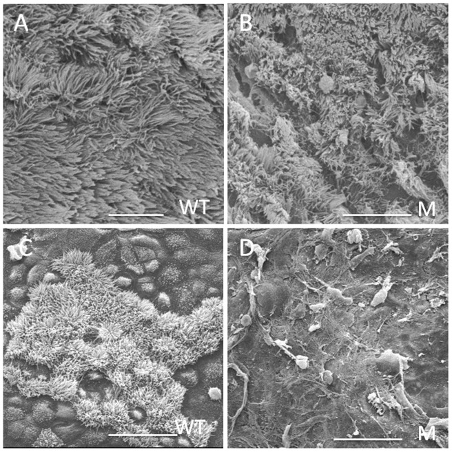
Figure 3. Analyses of cilia in the epithelial cells of the middle ear and Eustachian tubes.
(A) Normal-looking cilia in the middle ear epithelial cells of 25-day old wild-type mice, the cilia were well organized. (B) Cilia in the middle ear epithelial cells of 25-day old Spag6-deficient mice, notice that the cilia were largely disordered. Cilia were present in the promontorium tympani of 25-day old wild-type mice (C), but were prematurely lost in the Spag6-deficient mice (D). WT, wild-type; M, mutant; Bars: A, 10 µm; B, 15 µm; C and D, 25 µm.

