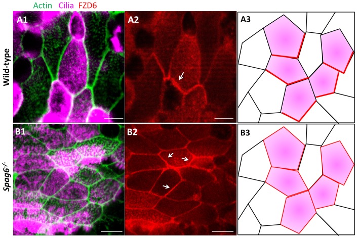
Figure 5. FZD6 protein polarity is lost in mutant tympanic ciliary epithelium.
(A–B) The distribution of the core PCP protein, FZD6, was examined by immunofluorescence staining. Ciliated epithelial cells were marked with anti-acetylated-α-tubulin antibody (rose). (A1, A2) In the wild-type mice (at P25), FZD6 protein localized asymmetrically to the cell cortex at the level of the apical junctions (arrow in A2). (B1, B2) However, in the mutant mice (at P25), FZD6 protein lost its polarized distribution and was located on the entire membrane at the apical level (arrows in B2).(A3, B3) Schematic diagram of FZD6 protein (red line) distribution in wild-type and Spag6−/− mice. The color codes are the same as that in panel A1 and B1. Bars: A, 5 µm; B, 7.5 µm.

