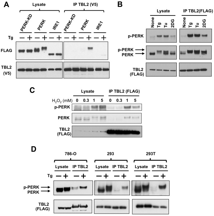
Figure 3. Preferential binding of TBL2 to phospho-PERK.
(A) 293T cells were transiently co-transfected with pTBL2 (V5-tag) and either pFLAG-PERK, pFLAG-PERK(K621A) or pFLAG-IRE1 and then were treated with 300 nM thapsigargin (Tg) for 2 h. The cell lysates were immunoprecipitated with anti-V5 antibody and immunoblotted with anti-FLAG or anti-V5 antibody. (B) 293T cells were transiently transfected with pFLAG-TBL2 and then were treated with 300 nM thapsigargin (Tg), 4 µg/ml tunicamycin (Tu) or 10 mM 2-deoxyglucose (2DG) for 2 h. Endogenous PERK protein was detected with anti-PERK or anti–phospho-PERK antibody. (C) 293T cells were transiently transfected with pFLAG-TBL2 and then were treated with the indicated doses of hydrogen peroxide (H2O2) for 4 hour. After immunoprecipitation with anti-FLAG antibody-conjugated beads, each protein was immunoblotted with the indicated antibody. (D) 786-O, 293 and 293T cells were transiently transfected with pFLAG-TBL2 and then were treated with 300 nM thapsigargin (Tg) for 1 hour. After immunoprecipitation with anti-FLAG antibody-conjugated beads, each protein was immunoblotted with the indicated antibody.

