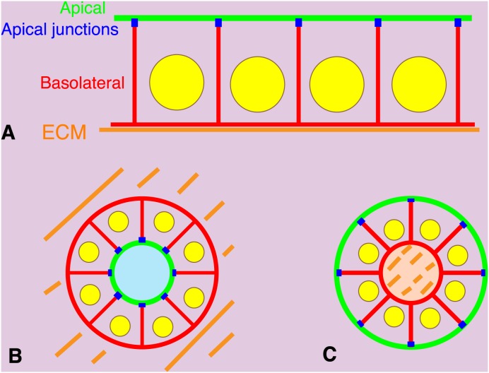
Figure 1. Apical-basal polarities of epithelial cells in 2-D or 3-D culture.
(A) Polarized epithelial cells in a 2-D sheet. Cells are on extracellular matrix (ECM, orange) coated artificially or deposited by the cells themselves. Plasma membranes facing the ECM or adjacent cells are called basolateral membranes (red). The remaining membrane areas are called apical membranes (green). Apical junctions (blue) are formed at the border between basolateral and apical membranes. (B) Polarized epithelial cells forming a spheroid in the ECM gel. Basolateral membranes are formed on the outside surface of the spheroid facing the ECM. Apical membranes are formed inside the spheroid. (C) Polarized epithelial cells forming a spheroid in suspension culture. Concentration of the ECM deposited by the cells themselves appears higher within the spheroid. Apical membranes are formed on the outside surface of the spheroid facing the culture medium. Basolateral membranes are formed on inside the spheroid.

