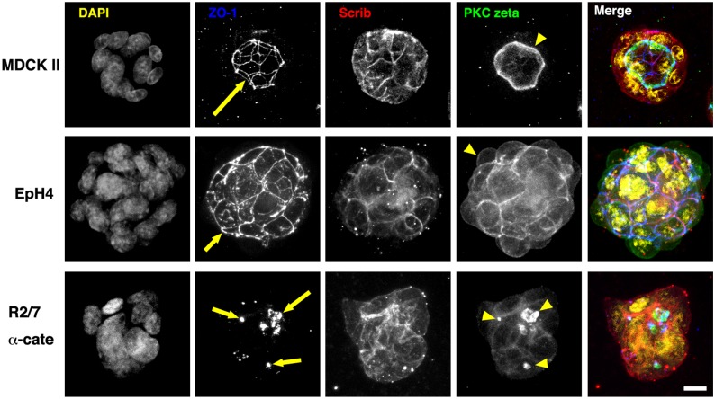
Figure 4. Apical-basal polarity and lumen formation of epithelial spheroids in collagen gels.
Spheroids of MDCK II, EpH4, and R2/7 α-cate cells after 3-day culture in type I collagen gels. Cells were stained for DNA (yellow), ZO-1 (blue), Scrib (red) and PKC zeta (green). Series of confocal optical sections were obtained and projected onto a single plane. In MDCK II cells, single lumens lined with PKC zeta (yellow arrowhead) and enclosed by ZO-1 network (yellow arrow) were observed. EpH4 cells had no lumen, and the ZO-1 network (yellow arrow) was distributed on the surface of the spheroid. PKC zeta also accumulated at the surface of the spheroid (yellow arrowhead). R2/7 α-Cate cells had several small lumens inside the spheroid (yellow arrows and arrowheads). Bar, 10 µm.

