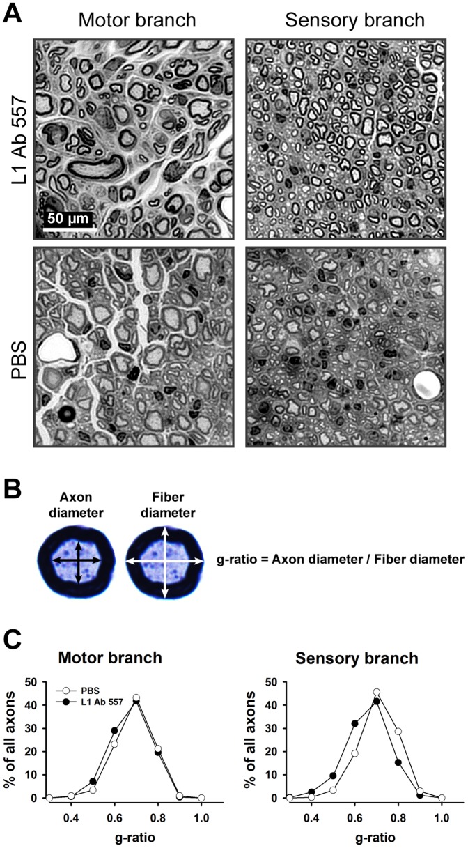
Figure 4. Analysis of myelination in regenerated femoral nerves in mice treated with L1 Ab 557 or PBS in the conduits applied to the transected nerves.
(A) Representative images of the motor and sensory nerve branches from L1 Ab 557 or PBS treated mice. (B) Mean orthogonal diameters of the axon (black arrows) and of the nerve fiber (white arrows) were measured and the degree of myelination was estimated by the ratio of axon to fiber diameter (g-ratio). (C) Normalized frequency distributions of g-ratios in regenerated motor and sensory nerve branches. Regenerated nerves were studied 12 weeks after injury. Ten mice per group were analyzed. The shift in distributions of g-ratios to the left in the group treated with L1 Ab 557 in the sensory nerve shows better myelination compared to the group of animals treated with PBS (p<0.05, Kolmogorov-Smirnov test).

