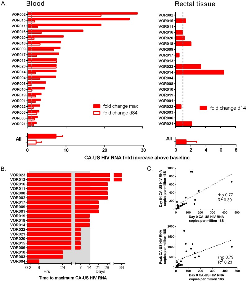
Figure 2. Individual changes in CA-US HIV RNA in blood and tissue.
A) Fold change in CA-US HIV RNA following vorinostat in CD4+ T-cells from blood (left panel) and rectal tissue (right panel) compared to baseline. The maximum fold change in CA-US HIV RNA on study (solid column) and change at day 84 (open column) is shown for CD4+ T-cells from blood; and change at day 14 for rectal tissue is shown for each participant (upper panel) and the median (IQR) change for all participants (lower panel). The grey dashed line indicates 1-fold change. B) Time to reach maximum fold increase in CA-US HIV RNA for each participant. Grey shaded box represents the time on vorinostat. (C) Correlation between baseline CA-US HIV RNA and peak CA-US HIV RNA (left panel) and day 84 CA-US HIV RNA (right panel).

