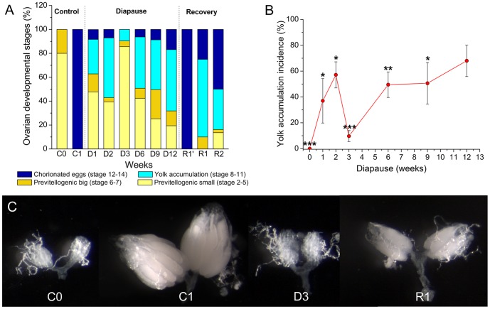
Figure 1. Ovarian development and yolk accumulation are arrested during diapause.
A Ovarian developmental stages (%) in virgin female flies Drosophila melanogaster Canton S (A), kept for 1–12 weeks at 11°C and short photoperiod 10L:14D (diapause, D1–D12) or after recovery for 1 week after 3 weeks of diapause (R1′), 1 (R1) or 2 weeks (R2) after 6 weeks of diapause. Recovery was carried out at 25°C and normal photoperiod 12L:12D, light/dark. As comparison we used 3–6 h old (newly eclosed) virgin flies (C0) or virgin flies kept under normal conditions for 1 week (C1). The developmental stage of ovaries was assessed as outlined in Material and methods. Note that one week control flies (C1) have fully developed ovaries. Data are presented as means ± S.E.M, n = 4 independent replicates with 8–12 flies in every replicate. B Yolk accumulation incidence (%) in ovaries of virgin female flies (Canton S) kept at the same conditions as in 1A. 1-week old flies, kept under normal conditions (C1) were used as reference. Yolk accumulation incidence (%) was defined as the presence yolk deposit even in a single oocyte (stage 8) up to formation of one/several chorionated eggs (stages 12–14) in mostly previtellogenic ovaries of diapausing flies. Data are presented as means ± S.E.M, n = 4 independent replicates with 8–12 flies in every replicate. We indicate data that are significantly different from the C1 flies kept for one week at normal conditions (C1) that display 100% yolk incidence (Student t-test with *p<0.05, **p<0.01, ***p<0.001). See Fig. S1 in File S1 for ovary development in dilp mutants and Fig. S6A, B in File S1 for w1118 flies. C Representative images of ovaries at selected stages of Canton S.

