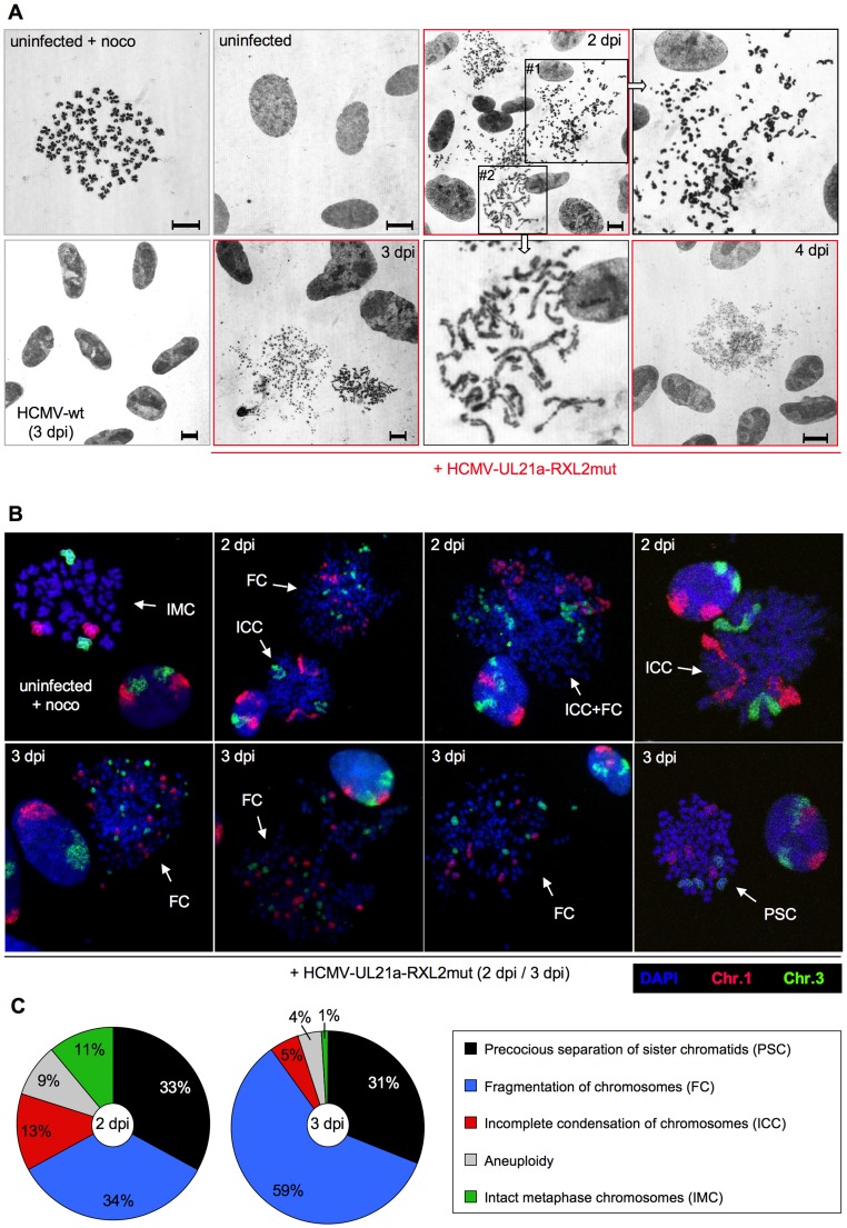
Figure 9. Precocious separation of sister chromatids and progressive chromosome fragmentation predominate the chromosomal appearance of HCMV-UL21a-RXL2mut-infected cells.
(A) Metaphase spreads from nocodazole-treated, non-infected cells were subjected to Giemsa staining and compared to equally prepared chromosomal material of HCMV-wt and HCMV-UL21a-RXL2mut-infected cells at 2 to 4 days post infection (dpi). Where indicated, magnified views of the encircled areas #1 and #2 in the adjacent image of RXL2mut-infected cells are shown. Scale bars: 5 µm. (B) Metaphase spreads were analyzed by fluorescence in situ hybridization (FISH) using whole chromosome painting probes for chromosomes 1 and 3. DNA was counterstained with DAPI. Typical examples of cells with intact metaphase chromosomes (IMC), fragmented chromosomes (FC), incomplete chromosome condensation (ICC) or precocious separation of sister chromatids (PSC) are shown and labeled accordingly. (C) Quantitative evaluation of FISH analysis based on at least 100 mitotic HCMV-UL21a-RXL2mut-infected cells per sample. Non-mitotic cells were not included in the analysis.

