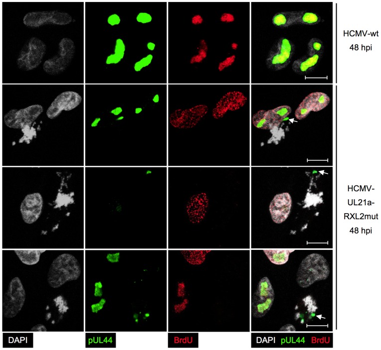
Figure 11. Impaired viral DNA replication in mitotic cells.
Cells were infected with HCMV-UL21a-RXL2mut and prepared at 48 hpi for immunofluorescence microscopy. Chromatin condensation (DAPI), localization of viral DNA replication compartments (pUL44) and DNA synthesis (BrdU) were analyzed and representative images are shown. The right column displays the merged fluorescence channels. Arrows point to the small pUL44-positive areas in metaphase cells. Scale bars: 5 µm.

