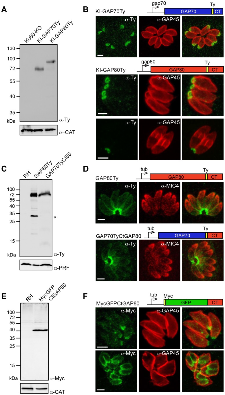
Figure 1. A family of GAPs anchored in different sub-compartments of the IMC.
A. Total lysates from Ku80-KO parasites expressing a Ty-tagged endogenous GAP70 or GAP80 (KI: knock-in) analyzed by western blot using anti-Ty antibodies and catalase (CAT) as loading control. B. Localization of KI-GAP70 at the apical cap and KI-GAP80 in a ring-like structure at the basal pole assessed in intracellular parasites using anti-Ty as well as anti-GAP45 or anti-IMC1 antibodies that stain the periphery or the IMC, respectively. Scale bars: 2 µm. C. Immuno-blot of total lysates of RH parasites expressing a second copy of GAP80Ty or GAP70TyCtGAP80 under the control of the tubulin (Tub) promoter. Profilin (PRF) was used as a loading control. The star indicates a degradation product. D. Localization of the second copy of GAP80Ty or GAP70TyCtGAP80 assessed in intracellular parasites using anti-Ty together with anti-MIC4 antibodies that stain the micronemes. Scale bars: 2 µm. E. Total lysates from RH parasites expressing MycGFPCtGAP80 under the control of the tubulin promoter analyzed by western blot using anti-Myc antibodies and CAT as a loading control. F. Localization of MycGFPCtGAP80 at the posterior sub-compartment of the IMC in mature parasites and in growing daughter cells. Scale bars: 2 µm.

