
Figure 4.
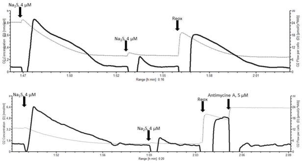
The plots show original tracing of two selective experiments conducted in order to show the effects of decreasing oxygen concentrations on sulfide turnover and thus elimination. The plots report the oxygen concentration (thin dotted line, left Y-axis), and the oxygen flux (bold closed line, right Y-axis) over time. The oxygen concentration was within the same range as for the sulfide titration experiments. For these experiments we used intact AMJ2-C11 cells in the FCCP-induced uncoupled state. 0.5 μM rotenone were added in order to inhibit mitochondrial respiration at the level of complex I. This was done in order ensure that any subsequent increase in oxygen flux following a sulfide injection would be attributable exclusively to sulphide turnover. Then we injected two sequential 4 μM boluses of the sulfide donor Na2S as indicated by the arrows. The first bolus induced a pronounced response in oxygen flux, whereas the response to the second bolus was much weaker. The oxygen concentrations were 4.9 μM (upper plot) and 3.4 μM (lower plot) before the first injection, and 1.6 (upper plot) and 1.1 (lower plot) before the second bolus. To exclude that a residual inhibition of the cytochrome-c oxidase was responsible for this weak response the medium was reoxygenated (Reox, see arrows), to increase the oxygen concentration to 3.8 μM in the experiment shown by the upper plot, and 5.3 μM in the lower plot. Under the higher oxygen concentrations the oxygen flux supported by sulfide immediately re-increased again to values approaching those after the injection of the first bolus. This suggest that the low oxygen flux observed after the second bolus of sulfide was likely due to the low oxygen concentration in the medium, in spite of a sufficient amount of sulfide available as substrate. In the experiment shown in the lower diagram, the complex III inhibitor antimycine A (5 μM) was injected after the final reoxygenation of the medium. This was done to show that after the inhibition of complex III the residual cellular respiration goes back to the values preceding any addition of sulfide. This indicates that the presence of sulfide in the chamber does not interfere with the oxygen sensors of the oxygraph, which could alternatively explain the increase in oxygen flux following the sulfide boluses.