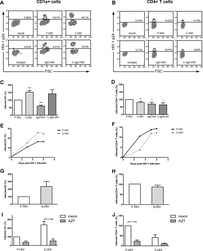
Figure 1.
Complement opsonization of HIV-1 enhances infection of DCs but decreases infection of CD4+ T cells. The cervical tissue biopsies were infected with different forms of HIV-1BaL, either free (F-HIV), complement opsonized (C-HIV), antibody opsonized (IgG-HIV), virions opsonized by a cocktail of complement and antibodies (C-Ig-HIV) or mock-treated, by spinning the cultures for 2 h at 37°C. The tissues were washed and transferred to six-well plates and cultured for 3–6 days. (A, B) The emigrating cells were harvested and stained with (A) anti-CD1a and anti-HIV-1 mAbs for DCs, and (B) anti-CD3, anti-CD4, and anti-HIV-1 mAbs for CD4+ T cells and the level of infection was assessed by flow cytometry. (C, D) The level of HIV-1 infection in the different (C) DC (N = 10–32) or (D) T-cell (N = 11–32) experiments was normalized with F-HIV set as 100%. (E, F) The kinetics of HIV-1 infection of emigrating (E) DCs and (F) CD4+ T cells were measured day 3–6. (G, H) The level of HIV-1 infection of emigrating (G) DCs and (H) T cells (N = 4–5) exposed to F-HIV or HIV-1 opsonized with seminal fluid was also assessed. (I, J) The level of HIV-1 infection of emigrating (I) DCs and (J) T cells from cervical tissue biopsies (N = 3) pre-exposed to mock or AZT followed by infection with F-HIV or C-HIV was measured on day 4. Mock or AZT was present throughout the whole 6-day culture. Statistical significance was tested using a two-sided paired t-test. *p < 0.05, **p < 0.001, ***p < 0.0001. Data are shown as mean + SEM of the indicated number of samples, each assessed in its own experiment.

