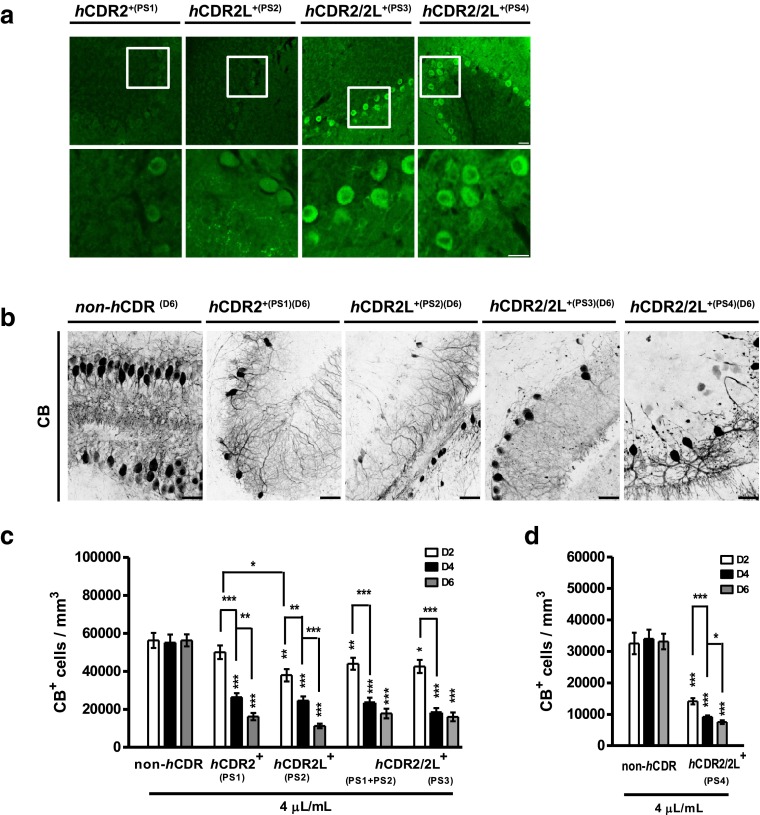
Fig. 2.
Calbindin D28K immunoreactivity loss in Purkinje cells in response to experimental induced human CDR antibody-mediated PCD. a Human CDR (hCDR)/PC binding was visualized by anti-human IgG AF488 staining of 8 μm cryostat rat cerebellum sections incubated with hCDR+ patients’ sera (1:2000). Magnification of the conducted cerebellar PC region micrographs (lower panel) shows that both hCDR2 and hCDR2L antibodies bound to the PC soma. Scale bars 25 μm. b Multiphoton micrographs of cOTSCs: Independent of the Ab target, all hCDR sera (4 μL/mL; 6 days; PS1-4) led to CB-positive (CB+) PC loss and altered their dentritic morphology; scale bars 40 μm. c, d Calculation of CB+ cells/mm3 after 2, 4, and 6 days of hCDR internalization revealed pathological effects of hCDR compared to non-hCDR control over time (nE = 4). Data in mean ± SEM. Non-parametric two-tailed paired Mann–Whitney’s U test. *p < 0.05; **p < 0.01; ***p < 0.001. The percentage changes to controls are summarized in Table 1

