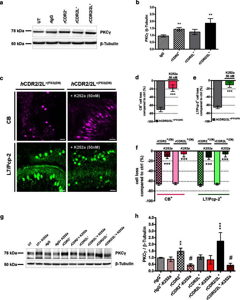
Fig. 7.
Protein kinase C gamma expression is up-regulated by CDR2 and CDR2/2L-Ab internalization. a Representative Western blot: PKCγ expression after rCDR internalization. b PKCγ expression was significantly increased after CDR2 and CDR2/2L internalization (125 ng/mL; 6 days; n E = 14). c Multiphoton micrographs demonstrate that the hCDR2/2L+(PS3)-induced loss of CB (magenta) and L7/Pcp-2 (green) was minimized by PKCγ antagonist K252a (50 nM) co-treatment; scale bars 40 μm. Stereological counting of CB+ and L7/Pcp-2+ PCs in the obtained micrographs supported the positive effect of K252a on CDR antibody-induced pathology by showing a loss of <10 % compared to control (hCDR2/2L+(PS3) [4 μL/mL] d CB: n E = 4; e L7/Pcp-2: n E = 4; f rCDR2 and rCDR2L [125 ng/mL] CB: n E = 6; L7/Pcp-2: n E = 6). g Representative Western blot: PKCγ expression after rCDR/K252a co-treatment. h K252a co-treatment reduces the rCDR-induced PKCγ expression rise in the rCDR2 and rCDR2/2L group (125 ng/mL; n E = 5) after K252a co-treatment. Investigated samples: 6 days of CDR internalization; data in mean ± SEM; non-parametric two-tailed paired Mann–Whitney’s U test. *p < 0.05; **p < 0.01; ***p < 0.001; # p < 0.003; Table 1: CDR antibody effects in percentage

