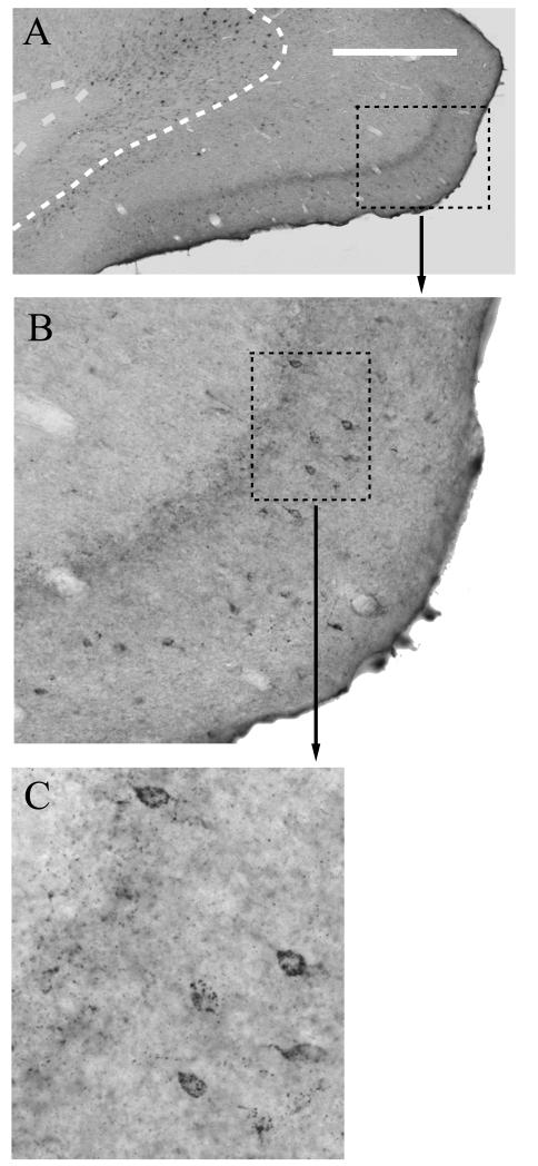
Figure 6.
Photomicrograph of caudal retrosplenial cortex showing the presence of additional (trans-neuronal) retrograde label in lamina II (see B,C) following extended survival times after a bilateral injection of wheat germ agglutin (WGA) into the anterior thalamic nuclei. A) Photomicrograph of caudal retrosplenial cortex (Rga, Rgb) reacted for WGA following a 3 day survival period. The dashed line marks the boundary between lamina V and VI. Scale bar = 500μm. B) Higher magnification photomicrograph of region indicated by a dashed box in (A), showing both anterograde label (laminae I-IV) and retrograde label concentrated in lamina II cells. C) Further high magnification photomicrograph of the retrograde label in the region indicated by the dashed box in B. Note, the occasional cell in lamina III/IV. All sections are in the coronal plane.

