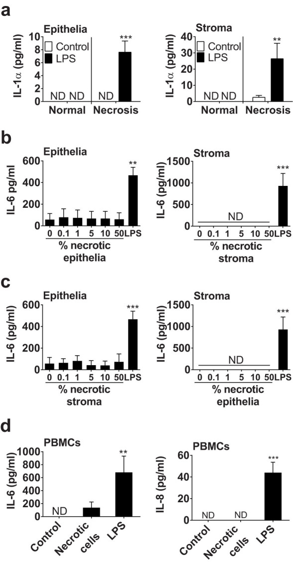
Figure 4. Necrotic cells do not simulate inflammation.

(a) Endometrial epithelial and stromal cells were cultured in control medium or medium containing 0.1 μg/ml LPS for 24 h prior to collection of supernatants from normal cells or from cells in which necrosis was induced by cycles of freezing and thawing. Data are mean + SEM, from 3 independent experiments. Data were analyzed by GLM ANOVA, using the Bonferroni post hoc multiple comparison test; values differ from control, *** p < 0.001, ** p < 0.01. (b, c) Endometrial cells were cultured for 24 h in media containing the indicated percent of a solution of necrotic (b) homologous or (c) heterologous cells, with medium containing 0.1 μg/ml LPS used as a positive control. Cell-free supernatants were collected and the concentrations of IL-6 were measured by ELISA. (d) PBMCs were treated with control medium, or media containing 10% necrotic stromal cells or 0.1 μg/ml LPS for 24 h. Cell-free supernatants were collected and IL-6 and IL-8 measured by ELISA. Data are mean + SEM from 4 independent experiments. Data were analyzed by GLM ANOVA, using the Dunnett's pairwise multiple comparison t-test; values differ from control, *** p < 0.001, ** p < 0.01, ND = not detected.
