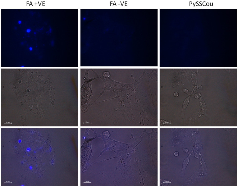
Figure 6. Fluorescence microscopy images of B16-F10 cells after co-incubation with FA+VE ZG-20 NPs, FA–VE ZG-20 NPs, and PySSCou molecule.
Upper three: Fluorescence images were taken in the DAPI (4′,6-diamidino-2-phenylindole) channel. Excitation filter: 350/50 nm and emission filter: 460/50 nm. Middle three: Bright field images of the respective cells. Bottom three: Merged images of the respective bright field and fluorescence cell images. Scale bar is 20 μm.

