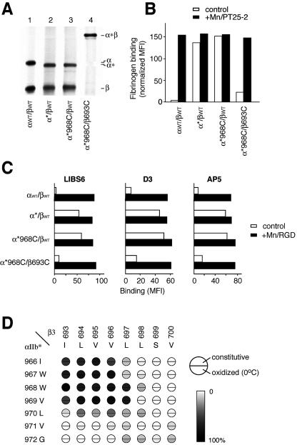
Figure 4. Formation of Intersubunit Disulfide Bonds in the TM Domain of αIIb*β3 and Effect on Ligand Binding and LIBS Epitopes.
(A) Immunoprecipitation. Immunoprecipitation of [35S]-labeled receptors and nonreducing SDS-PAGE and fluorography was as described in Figure 2.
(B) FITC-fibrinogen binding. Binding was determined by immunofluorescence as described in Figure 3.
(C) LIBS exposure. Three different anti-LIBS mAbs (LIBS6, D3, and AP5) were used to probe the conformational state. mAb binding is expressed as the mean fluorescence intensity in the absence (control, open bars) or presence (+Mn/RGD, black bars) of Mn2+ and RGD peptide.
(D) Disulfide bond formation efficiency. Disulfide bond formation in αIIb*β3 heterodimers with the indicated residues mutated to cysteine was determined as described in Figure 2B.

