
Figure 3.
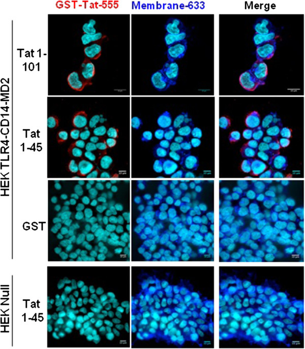
Analysis of Tat and- TLR4-MD2 labelling at the cell surface. HEK cells null or HEK-TLR4-MD2-CD14 were pre-incubated with GST-Tat 1–101 or GST-Tat 1–45 or control GST during 15 min. GST+/−Tat were labelled with an anti-GST antibodies (red). WGA-633 (blue) was used to label the membrane of cells and DAPI (cyan) was used as a nuclear marker. Scale bars are represented right down of each images.