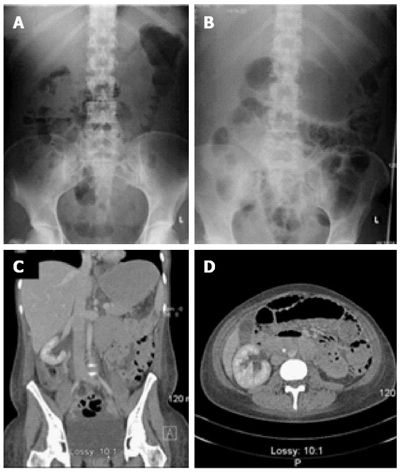
Figure 2.

Radiology of case two. (A) and (B) comparative pre and postoperative plain radiology showing new and extensive intramural gas in the latter; C and D: Axial computerized tomograms taken on the 8th postoperative day showing marked pneumatosis intestinalis.
