
Figure 1.
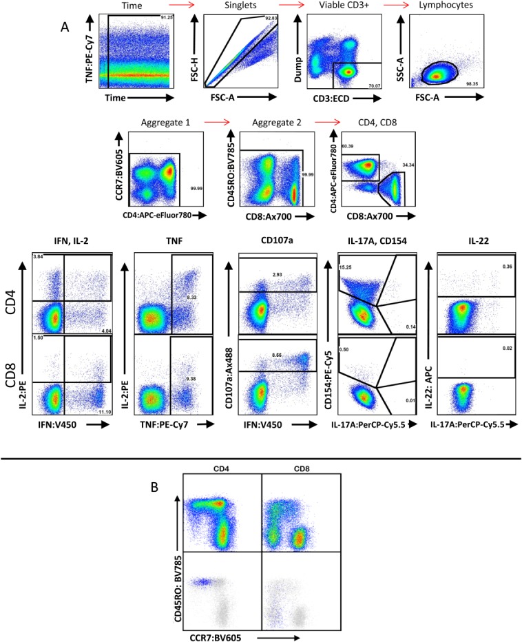
Example staining of adult human PBMC following stimulation. (A) The first two rows demonstrate the gating hierarchy from total sample to CD4/CD8 identification. A time gate is used to exclude pressure aberrations from the cytometer that may have occurred during sample acquisition. Aggregate gates are used to exclude brightly positive events that may result from antibody or cell aggregation. The bottom row demonstrates gating for cytokines and functions from CD4+ (top half) and CD8+ (bottom half) events resulting from stimulation with Staphylococcal enterotoxin B. Note that IL-17A and CD154 were gated on the same plot to avoid mischaracterization resulting from the increased spectral overlap observed from their fluorochromes. (B) Example plots showing memory profile of CCR7 versus CD45RO in CD4+ and CD8+ populations. Using CMV pp65 peptide pool-stimulated PBMC from a CMV-reactive donor, IFN-γ+ events (blue) were overlaid onto these plots (gray), confirming localization of these events to the effector memory and effector compartments.