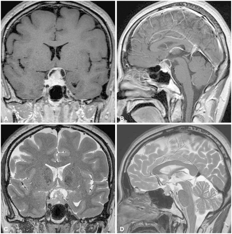
Fig. 1.
Preoperative magnetic resonance imaging shows a heterogeneously enhanced mass involving the sella in a T1-weighted gadolinium-enhanced coronal (A) and sagittal view (B). T2-weighted coronal (C) and sagittal (D) images reveals solid and cystic components of the tumor with compression of an optic apparatus.

