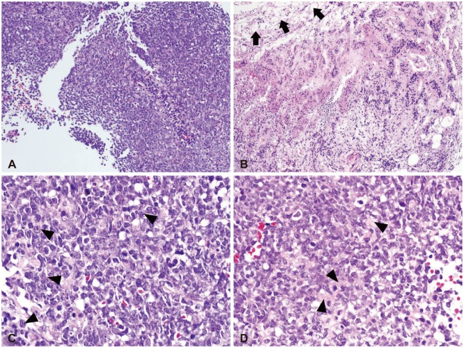
Fig. 2.
Low power views show relatively homogenous population of tumor cells with very high cellularity (A). At the inferior margin of the tumor mass, pituitary glandular structure (black arrows) is seen (B). High power views demonstrate a few tumor cells showing typical rhabdoid features with eccentric nuclei and prominent nucleoli (black arrowheads) (C). Abundant eosinophilic cytoplasm (black arrowheads) is noted (D). H&E, original magnification ×100 (A and B), ×400 (C and D).

