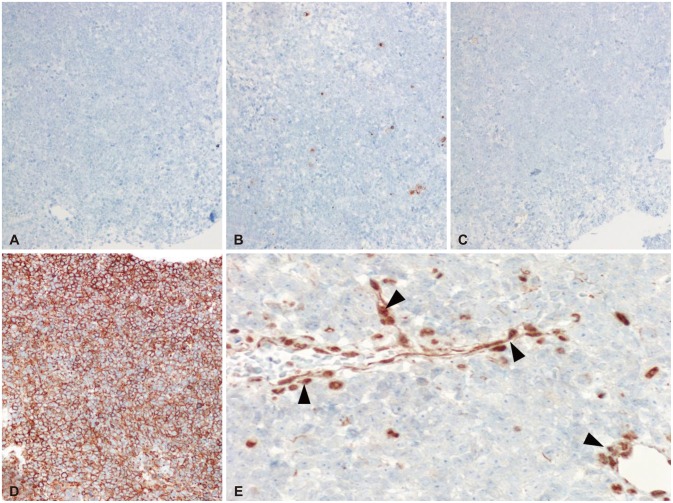
Fig. 3.
Immunohistochemistry (IHC) was negative for CD20 (A), cytokeratin (B), synaptophysin (C), and strong positive for CD99 (D). IHC stain for INI1 is negative in tumor cells whereas it is positive for endothelial cells (black arrowheads) (E). CD20-IHC, original magnification ×200 (A), cytokeratin-IHC, ×200 (B), synaptophysin-IHC, ×200 (C), CD99-IHC, ×200 (D), and INI1-IHC, ×400 (E).

