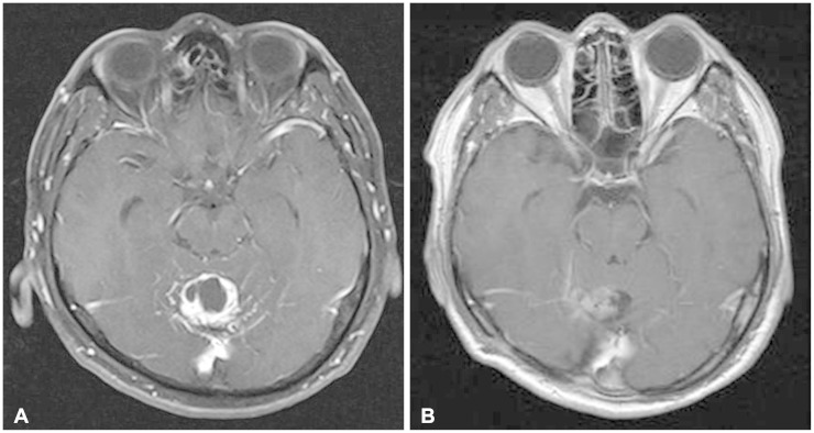
Fig. 1.
Pre and postoperative magnetic resonance images (MRIs). A: Preoperative gadolinium-enhanced T1-weighted MRI shows strongly enhancing mass involving the cerebellar vermis. B: Postoperative gadolinium-enhanced T1-weighted MRI. Tumor mass is completely removed and postoperative changes are observed.

