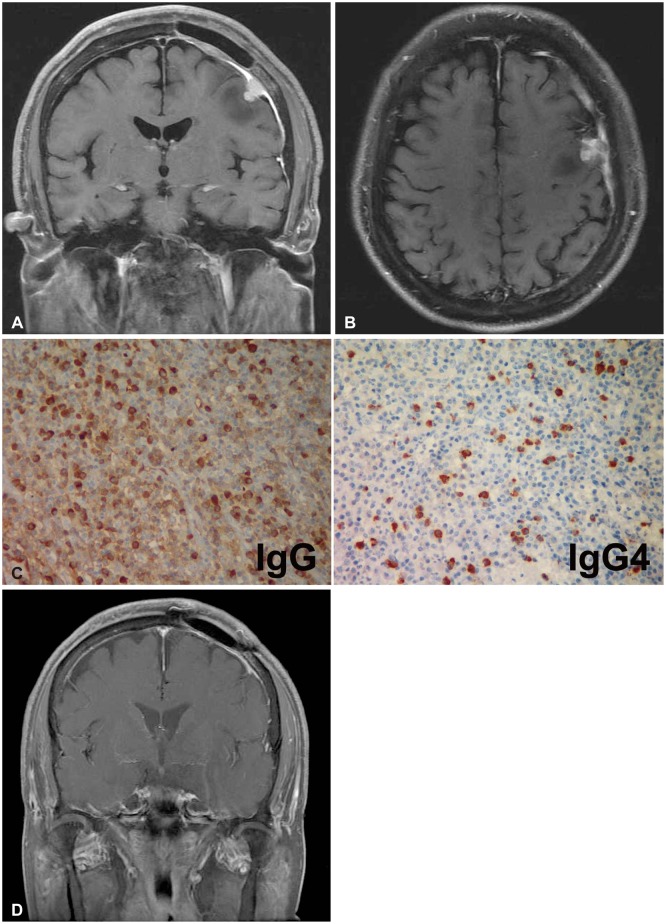
Fig. 3.
Postoperative magnetic resonance (MR) imaging and immunohistochemical staining. A and B: Eight months after surgery, contrast enhanced MR images showing the newly enhanced lesion adjacent to the previous mass. C: Immunohistochemical staining of immunoglobulin G (IgG) (left) and immunoglobulin G4 (IgG4) (right) (×400). D: Three months after immunosuppressant medication, brain MR imaging demonstrating that the enhanced mass and thickened dura had disappeared.

