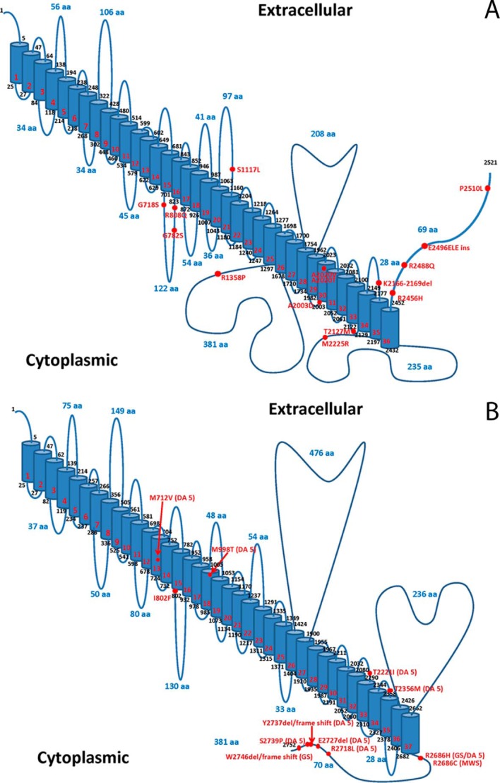
FIGURE 1.
Models of human PIEZO1 and PIEZO2. A and B, predicted membrane topology models of PIEZO1 (panel A, UniProt accession number Q92508) and PIEZO2 (panel B, UniProt accession number Q9H5I5) were created using Swiss-Prot prediction tools and the methodology of Eisenberg et al. (74). The locations of PIEZO1 mutations identified in hereditary xerocytosis (A) and PIEZO2 mutations identified in DA5, GS, and MWS (B) are marked. aa, amino acids.

