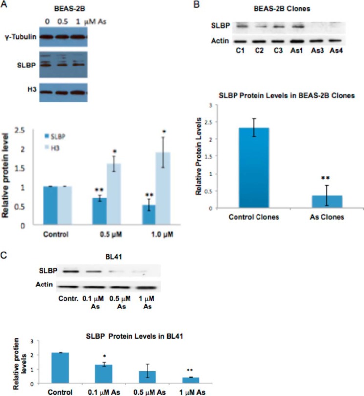
FIGURE 4.
SLBP protein levels are decreased by arsenic treatment. BEAS-2B and BL41 cells were treated with 0, 0.1, 0.5, or 1 μMNaAsO2 for 48 h, and BEAS-2B clones were transformed with NaAsO2. Cells were lysed with radioimmune precipitation assay buffer, and whole-cell lysate was run on 12% SDS acrylamide gels. A, representative Western blot of SLBP and H3 protein levels in BEAS-2B cells. Band intensities were quantified using Image J software. B, representative Western blot of SLBP protein levels in arsenic-transformed BEAS-2B clones and spontaneous clones. Band intensities were quantified using ImageJ software. Band intensities were pooled for control clones and arsenic-transformed clones to better visualize the dramatic decrease of SLBP levels in arsenic-transformed clones. C, representative Western blot of SLBP protein levels in BL41 cells. Band intensities were quantified using ImageJ software. Statistical significance was calculated using an unpaired, two-tailed t test with * indicating a p value less than 0.05 and ** indicating a p value less than 0.01. Error bars represent S.D. Contr., control.

