
FIGURE 6.
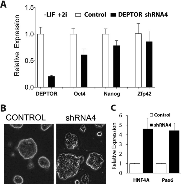
Validation of DEPTOR knockdown-mediated mESC differentiation. A, R1 mESCs were infected with control or DEPTOR shRNA4, and cells were selected using puromycin. Cells were then grown in the absence of LIF, mRNA was extracted, and qPCR was performed for Deptor, Oct4, Nanog, and Zfp42 as in Fig. 1. Actin was used as an internal control. B, morphological change of R1 mESCs upon DEPTOR knockdown in A was analyzed under phase contrast, and representative images are shown. C, Hnf4A and Pax6 mRNA expression mESCs after knockdown of DEPTOR using shRNA4. Error bars, S.E.