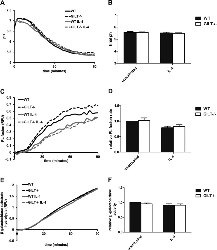
FIGURE 4.
Phagosomal maturation is not altered in the absence of GILT. BMMØs derived from WT and GILT−/− mice were incubated for 40 h in the presence/absence of 10 ng/ml IL-4. A, phagosomal pH calculated from excitation ratios of the pH-sensitive carboxyfluorescein-SE conjugated to experimental particles by regression to a standard curve. B, average final pH as measured 60 min post experimental particle internalization over 3 independent experiments. C and D, PL fusion as measured by FRET between phagocytosed Alexa Fluor 488-conjugated experimental particles and Alexa Fluor 594 hydrazide-pulsed lysosomes. E and F, phagosomal hydrolysis of a fluorogenic β-galactosidase substrate relative to a calibration fluor. A, C, and E, representative real-time traces. Relative fluorescence units (RFU) values are proportional to the degree of phagosome/lysosome fusion/β-galactosidase substrate hydrolysis. D and F, averaged rates relative to unactivated/untreated WT controls from three independent experiments. Relative rates were calculated between 40 and 60 min after particle internalization. Error bars denote S.E.

