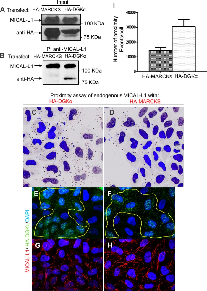
FIGURE 5.
DGKα forms a complex with MICAL-L1. DGKα and MICAL-L1 co-immunoprecipitate. A, HA-MARCKS (negative control) and HA-DGKα-transfected cells were lysed and blotted with anti-HA. B, in the same lysates, endogenous MICAL-L1 was pulled-down with anti-MICAL-L1 and eluted. Eluates were immunoblotted with anti-HA and anti-MICAL-L1. C–H, micrographs depicted show representative data from proximity ligation assays. Cells grown on coverslips were transfected with GFP-DGKα or HA-MARCKS (negative control). Duolink (proximity ligation) assay was performed using mouse anti-MICAL-L1 and rabbit anti-GFP. Dark dots in C–D indicate proximity (<40 nm) between MICAL-L1 and the HA-tagged protein. E–F, the same cells were also immunostained with anti-HA, to show similar degrees of transfection. G and H, endogenous MICAL-L1 was immunostained to display TRE morphology. I, number of proximity ligation events from 3 independent experiments (as done in C–D) was counted by Image J and plotted with standard deviation. Bar, 10 μm.

